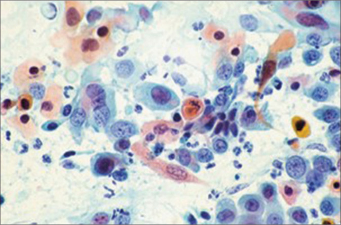
Figure 16:

Keratinizing HSIL cells showing pyknotic and hyperchromatic nuclei with high N: C ratio with coarse and irregularly distributed chromatin with attenuated cytoplasm. The numerous bizarre keratinizing (orange) cells and the granular background are highly suggestive of invasion. Image from The Bethesda System for reporting cervical cytology.
