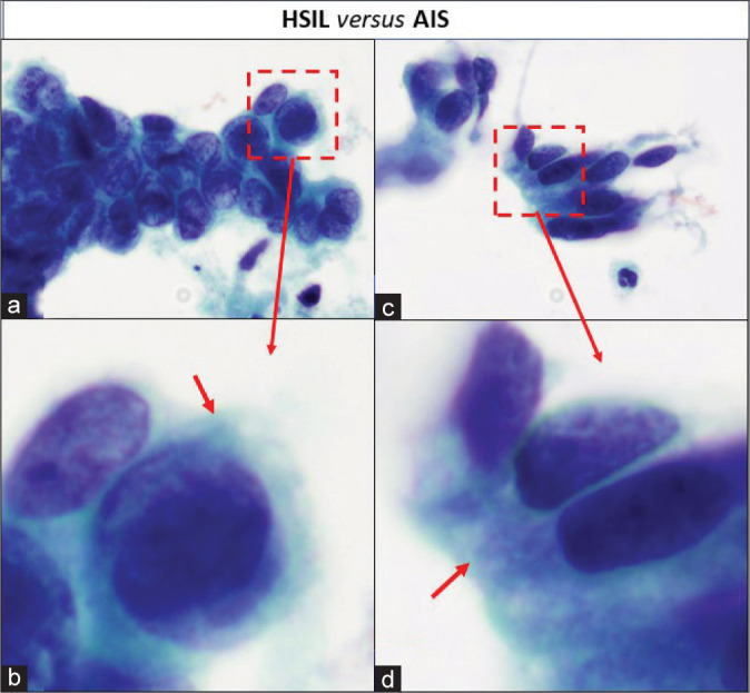
Figure 19:

Cytoplasm of HSIL versus AIS cells: (a and b) Groups of HSIL cells. The metaplastic cytoplasm of cells as seen in the cells at the periphery (arrow) is dense and homogeneous. (c and d) Groups of AIS cells. The cytoplasm shows lacy appearance (arrow).
