
Figure 24:
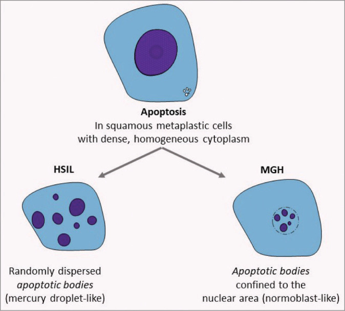
Apoptosis pattern in HSIL (CIN2) versus microglandular hyperplasia (MGH). Single-cell apoptosis in HSIL is not uncommon and typically shows dispersed nuclear fragments in the cytoplasm. In contrast, MGH cells which can also show single-cell apoptosis, show the apoptotic bodies restricted in the area of nucleus similar that observed in the normoblasts.