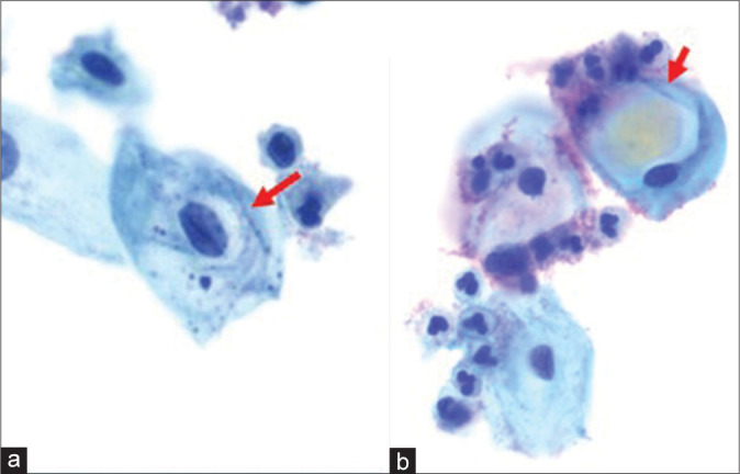
Figure 7:

(a) Pseudokoilocytes “tight halos” (long arrow) are common findings in atrophic specimens and with infections (e.g., Candida). (b) Glycogenated “navicular” cells (short arrow) also show perinuclear space occupied by yellow tinged “glycogen,” however, nuclei are negative for dysplastic features.
