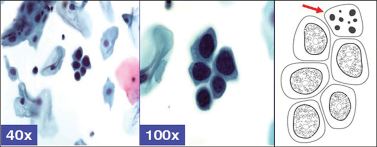
Figure 9:
Checkerboard pattern of HSIL, showing dark nuclei, smudgy chromatin, nucleoli, randomly distributed apoptotic bodies (arrow) (in contrast to MGH with apoptosis in the area corresponding with nucleus). This pattern correlates with CIN2 on biopsy. Adopted from CytoJournal open access. Chivukula M, Shidham VB. Cytojournal 2006;3:14.

