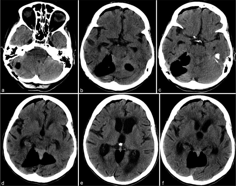
Figure 1:
Tension pneumocephalus after repeat microvascular decompression. (a-f) Respectively, caudal to rostral serial computed tomography images of the head without contrast material obtained preoperatively showing significant pneumocephalus in the posterior fossa in the previous microvascular decompression surgical corridor as well as the bilateral inferior frontal areas.

