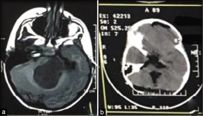
Figure 2:

(a) Preoperative MRI showing a tumor originating from the floor of the fourth ventricle filling the fourth ventricle and the lateral extension filling the cerebellomedullary cistern. (b) Postoperative CT after near-total excision through the telovelar approach with extensive opening of the cerebellomedullary fissure.
