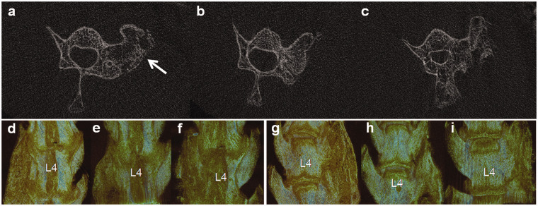
Figure 4.
μCT assessment of the fusion mass at four weeks after implantation (a, b, c Cross-section views, d–i 3D reconstructions). In the cross-section view, more new bone formation was induced in the 18 rats implanted with CBD-BMP2 + PRP (a) and the 18 rats with CBD-BMP2 (b) than the 17 rats with PRP (c). The white arrow indicates implant site. In 3D reconstructions images (d, e, f dorsal view, g, h, i ventral view), continuous and considerable fusion mass was only observed in rats implanted with CBD-BMP2 + PRP (d, g). In rats implanted with CBD-BMP2 (e, h), the fusion mass was relatively smaller than those with CBD-BMP2 + PRP (d, g). In rats implanted with PRP (e, h), the new bone formation was scattered, which was not merged into a whole mass. (A color version of this figure is available in the online journal.)

