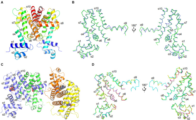
FIGURE 3.
Crystal structures of AbTetR and the minocycline bound AbTetR-Gln116Ala variant. (A) Crystal structure of TetR of A. baumannii AYE (rainbow color, N terminus: blue, C terminus: red). (B) Superimposition of AbTetR-A (green) and AbTetR-B (blue). (C) Binary structure of two dimeric Gln116Ala variants in complex with minocycline. Minocycline molecules are depicted as spheres (carbon: black; oxygen: red; nitrogen: blue). (D) Superimposition of the four monomers of minocycline-bound Gln116Ala (Gln116Ala-A: green, Gln116Ala-B: cyan, Gln116Ala-C: magenta, Gln116Ala-D: yellow).

