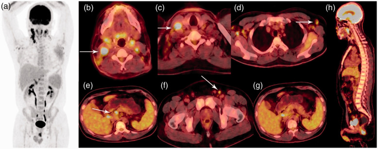
Figure 1.
Whole-body PET/CT scan findings. (a) The MIP image showed intense accumulation of FDG in the lymph nodes. (b)–(f) PET/CT fusion images showed multiple areas of hypermetabolic lymphadenopathy in the bilateral cervical, right supraclavicular, bilateral axillary, abdominal, and bilateral inguinal lymph nodes (white arrowhead). (g), (h) PET/CT fusion images showed mild, homogeneous FDG uptake in the spleen and bone marrow of the vertebral bodies.

