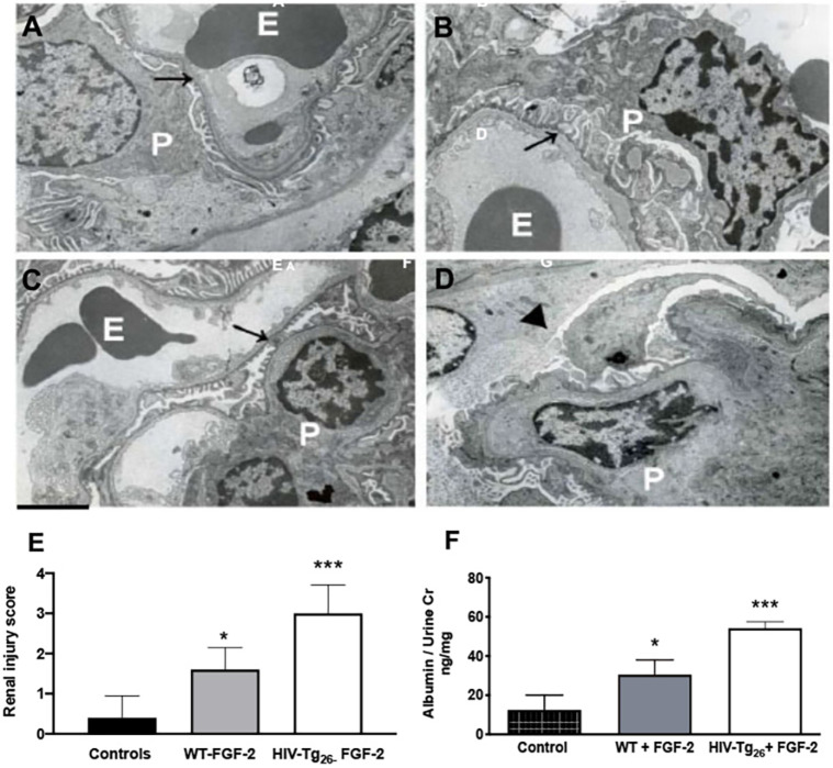
Fig. 2.
Circulating FGF-2 induced glomerular ultrastructural lesions in WT and HIV-Tg26 mice. A-D show representative electron microscopy pictures of renal glomeruli of WT and HIV-Tg26 mice injected daily intraperitoneally with either PBS control or recombinant human (rh) FGF-2 (10 μg/mouse/day×7 days). (A) Representative picture of normal glomerular endothelial cells and podocytes (arrow) from a WT mouse injected with PBS. E, erythrocytes; P, podocytes. (B) Representative picture of enlarged podocytes (arrow) and foot process effacement in an HIV-Tg26 mouse with HIVAN. (C) Representative picture of endothelial swelling (arrow) and mild podocyte foot process effacement from a WT mouse injected with rh-FGF-2. (D) Representative picture of a collapsed glomerular capillary (arrowhead), swollen endothelial cells and foot process effacement in an HIV-Tg26 mouse injected with rh-FGF-2. Scale bar: 2 μm (n=5 mice per group). (E) Renal injury scores generated as described in the Materials and Methods. WT mice injected with PBS served as controls. (F) Albuminuria in WT and HIV-Tg26 mice injected with rh-FGF-2. WT mice injected with PBS served as controls. The bars reflect the median and 95% confidence interval (CI) of five mice per group. ***P<0.001 and *P<0.05, compared to controls; one-way ANOVA (n=5 mice per group).

