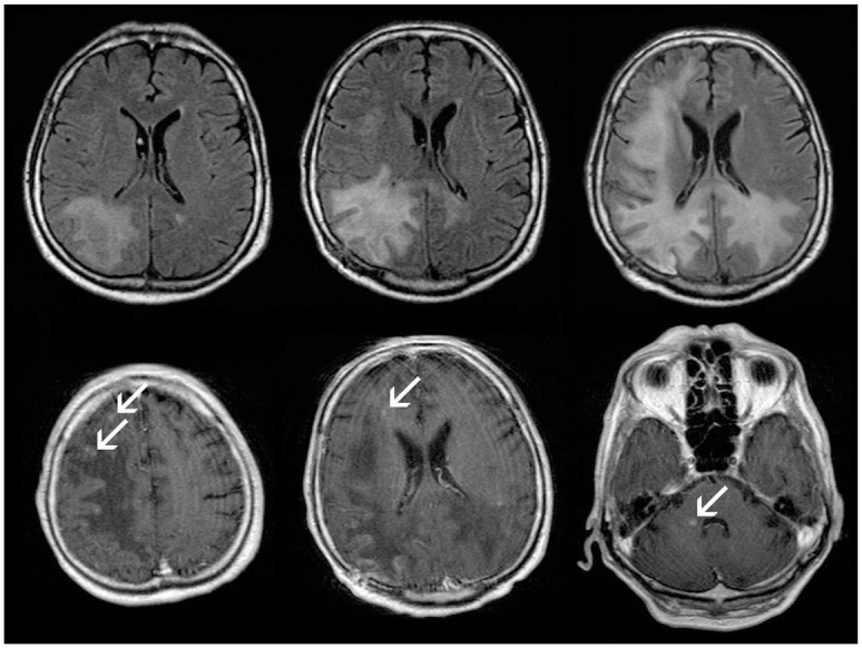
Figure 2.

Follow-up (within 2 months) axial T2-FLAIR (upper row) and axial contrast enhanced T1-SE (lower row) in patient 2, extensive progression of posterior accentuated, mostly subcortical PML-lesions is observed (upper row). Approximately 3 months after onset of the neurological symptoms, punctuate contrast enhancement (arrows) appear at the borders of the PML lesions suggesting the development of IRIS, corresponding to further clinical deterioration (lower row).
IRIS, immune reconstitution inflammatory syndrome; PML, progressive multifocal leukoencephalopathy.
