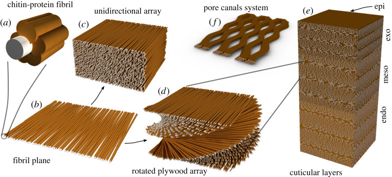
Figure 1.
A schematic diagram of the hierarchical structure of spider cuticle. (a) The chitin–protein basic building unit. The grey core represents the chitin crystal, whereas the proteins are represented in brown. (b) The fibril sheets. Chitin–protein fibrils are arranged roughly in parallel. (c) Unidirectional arrangement of fibril sheets. (d) Helicoidal arrangement of fibril planes. (e) The cuticle layers made of layers of parallel and helicoidal arrays. (f) Pore-canals cross-sections, the channels transverse the entire thickness of the procuticle. (Online version in colour.)

