Fig. 5.
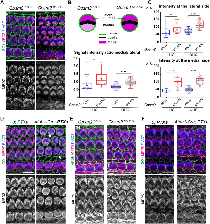
GNAI-GPSM2 promotes planar polarization of MPDZ and MPP5 at the apical membrane. (A) MPDZ labeling (magenta) at P4 (cochlear apex). MPDZ lacks higher enrichment at the lateral bare zone in the absence of GPSM2. (B,C) MPDZ enrichment in HCs at P4 (cochlear apex). The ratio between the medial and lateral (bare zone) MPDZ signals (B), and the individual signals at the bare zone (C, top) and medial apex (C, bottom) are plotted. Note how MPDZ signals increase in both compartments in Gpsm2 mutants, but more strongly at the medial apex. For all graphs: MpdzDEL/+ N=3, n=13 IHC, n=52 OHC; MpdzDEL/DEL N=3, n=18 IHC, n=62 OHC (Mann–Whitney U-test; (B) IHC: **P=0.0014, OHC: ****P<0.0001; (C) lateral IHC: **P=0.0064, lateral OHC: ****P<0.0001; medial IHC and OHC: ****P<0.0001). (D) MPDZ labeling (magenta) in Pertussis toxin (PTXa)-expressing HCs at P4 (cochlear apex). MPDZ lacks higher enrichment at the bare zone when GNAI is inactivated by PTXa. GNAI inactivation provokes graded OHCs misorientation so that the bare zone region (arrows) is not consistently lateral in PTXa OHCs. (E,F) MPP5 labeling (magenta) in GpsmDEL/DEL (E) and PTXa (F) HCs at P4 (cochlear apex). Like MPDZ (A-D), MPP5 is more uniformly enriched across the apical membrane when GPSM2 or GNAI is inactivated. In E, afadin (AFDN) was used instead of ZO1 to label the apical junctions. Scale bars: 10 µm.

