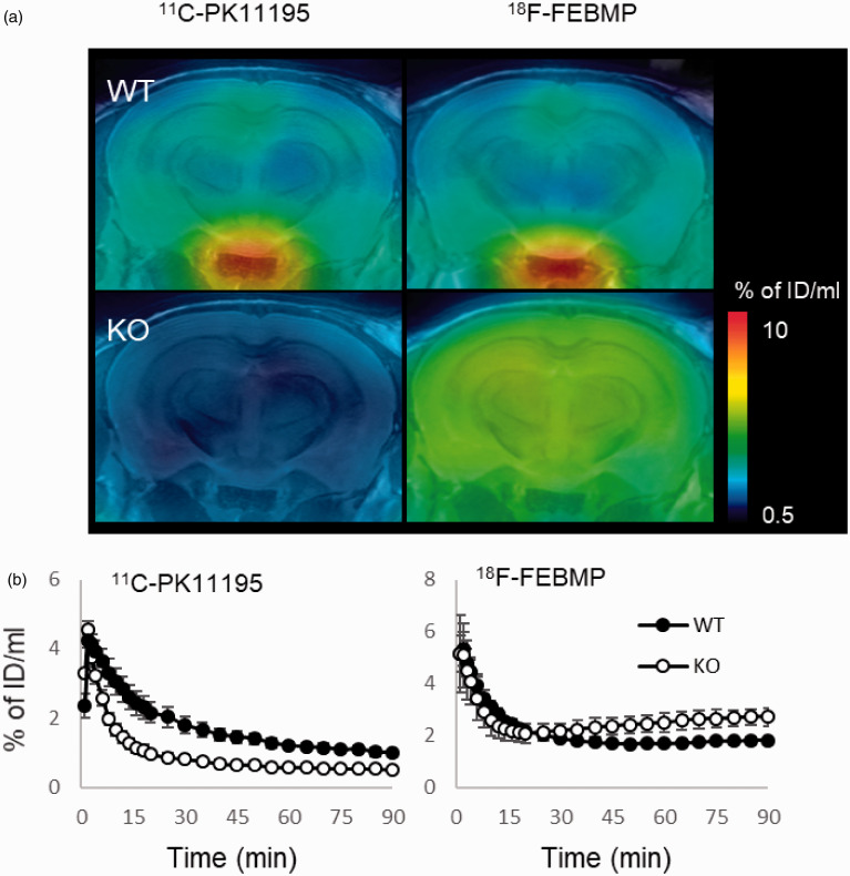
Figure 1.
In-vivo PET images of brains of WT and TSPO-KO mice following intravenous injection of TSPO radioligands. A: The representative 11C-PK11195 and 18F-FEBMP PET images of coronal mouse brain sections (2.4 mm posterior to the bregma) containing the neocortex and hippocampus in WT (male, 2-month old) and TSPO-KO (male, 2-month old) mice. PET images generated from averaged dynamic data (30–60 min) are overlapped with the MRI template of mouse coronal brain sections. B: Brain uptake was expressed as percentage of injected dose per ml brain volume in hippocampi from WT and TSPO-KO mice (each group included four 2-month-old male mice) over the scan time. The same individuals were used for 11C-PK11195 and 18F-FEBMP scans. Error bars represent standard deviation (SD). WT: wild-type, KO: TSPO-KO.

