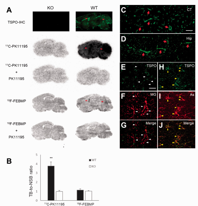
Figure 2.
Origin of TSPO expression and binding of radioligands in normal mouse brain. A: Immunohistochemistry with commercially available (C.A.) anti-TSPO antibody and representative in-vitro autoradiographic images of 11C-PK11195 and 18F-FEBMP in sagittal mouse brain sections from TSPO-KO (left) and WT (right) mice. Red arrowheads indicate lateral ventricles and cerebellums. B: Quantitative analysis for in-vitro binding. Data were from panel A. Total binding (TB) and non-specific binding (NSB) were calculated in the absence and presence of non-radiolabeled PK11195, respectively. Brain sections from six TSPO-KO and six WT mice were used for autoradiography with 11C-PK11195, and brain sections of three mice among the six TSPO-KO and WT mice, respectively, were used for autoradiography with 18F-FEBMP. There were significant main effects of genotype and tracer on in-vitro binding, [F(1, 14) = 83.92 and 105.7, respectively, p < 0.0001 by two-way ANOVA]. TB-to-NSB ratio of 11C-PK11195 in WT brain was significantly higher than in other three groups (**, p < 0.01 by post hoc Bonferroni’s test), while that of 18F-FEBMP in WT brain was similar to that in KO mouse groups (p > 0.99 by post hoc Bonferroni’s test). Error bars represent SD. C, D: Immunohistochemical analysis for TSPO origin. Most immunoreactivity of anti-TSPO antibody was from blood vessels (arrows in C and D) as shown in representative images of cortical and hippocampal regions. E-J: Double-channel photomicrographs of immunostaining of TSPO (E, H, and green in merged images G, J) and either microglia (MG; F and red in merged image G) or astrocytes (As; I and red in merged image J) demonstrated that resident astrocytes, but not microglia, express detectable TSPO in normal mouse brain. Scale bars: 50 μm (C, D); 20 μm (E-J).

