Figure 3.
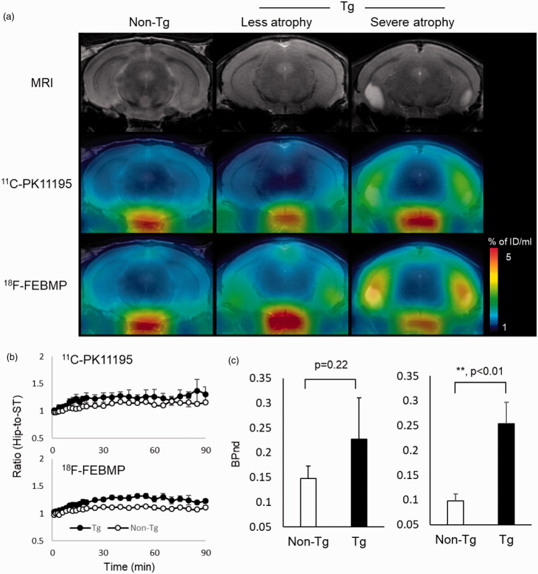
Tau lesion-associated microglial TSPO was more sensitively captured by in-vivo PET imaging with 18F-FEBMP than 11C-PK11195. A: T2 MRI images (upper row) and PET images with 18F-FEBMP (middle row) and 11C-PK11195 (lower row) of coronal mouse brain sections (2.8 mm posterior to the bregma) containing the neocortex and hippocampus in a male non-Tg (left column), and male PS19 Tg individuals with less (middle column) and severe (right column) brain atrophy at 9 months. The PET images generated from averaged dynamic data (30–60 min) are overlapped with the respective MRI brain templates (adult C57BL/6J) of coronal brain sections of individual mice. The images of middle (less atrophy) and right (severe atrophy) columns were from the same individuals, respectively. B: Time course of hippocampus (Hip)-to-striatum (ST) ratios of radioactivity of 11C-PK11195 (upper) and 18F-FEBMP (lower) C: Binding potential (BPnd) calculated by simplified reference tissue model with ST as reference tissue showing in-vivo binding of 18F-FEBMP (right), but not 11C-PK11195 (left), was significantly increased in Tg compared with non-Tg mice (n = 3 and n = 4 for 8-month-old male PS19 and age-matched non-Tg mice, respectively; t = −3.868, *, p < 0.05 by independent t-test for 18F-FEBMP). The same individuals were used for in vivo imaging of 11C-PK11195 and 18F-FEBMP. Error bars represent SD.

