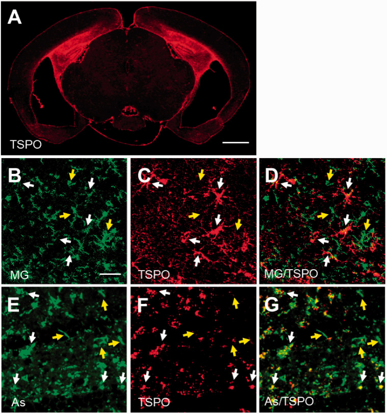
Figure 4.
Immunohistochemical analysis for TSPO origin in PS19 mouse. The immunoreactivity of anti-TSPO antibody was greatly increased in hippocampus of Tg mouse individual with overt brain atrophy (A), and double-channel photomicrographs of immunostaining of either microglia (MG; B and green in merged image D) or astrocytes (As; E and green in merged image G) and TSPO (C, F and red in merged images D, G) demonstrated that the majority of TSPO was activated microglia (white arrows in B-D) and astrocytes (white arrows in E-G), although there was still a subset of microglia and astrocytes lacking overt TSPO expression (yellow arrows in B-G) in lesioned hippocampus of Tg mouse. A part of TSPO-positive astrocytes seemed to be blood vessel-associated subtype morphologically. Scale bars: 1 mm (A); 20 μm (B-G).

