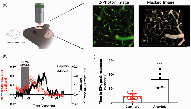
Figure 1.
Capillary response to whisker stimulus precedes arteriolar response. Capillary and arteriolar responses to whisker stimulation were evaluated using in-vivo two-photon microscopy in WT mice injected with 200 kD FITC-dextran to visualize vasculature: (a) schematic of in-vivo experimental set-up with representative two-photon and subsequent masked images; (b) red blood cell flux (capillaries) and red blood cell velocity (arterioles) in response to 10-s whisker stimulus. Magnitude change was calculated from the pre-stimulus baseline average, and data aligned by the 10-s stimulus timeframe; (c) time to 50% peak response following whisker stimulation is shorter in capillaries than arterioles (unpaired t-test, ****P < 0.0001). Data represent mean ± SD (n = 12 capillaries, n = 5 arterioles).

