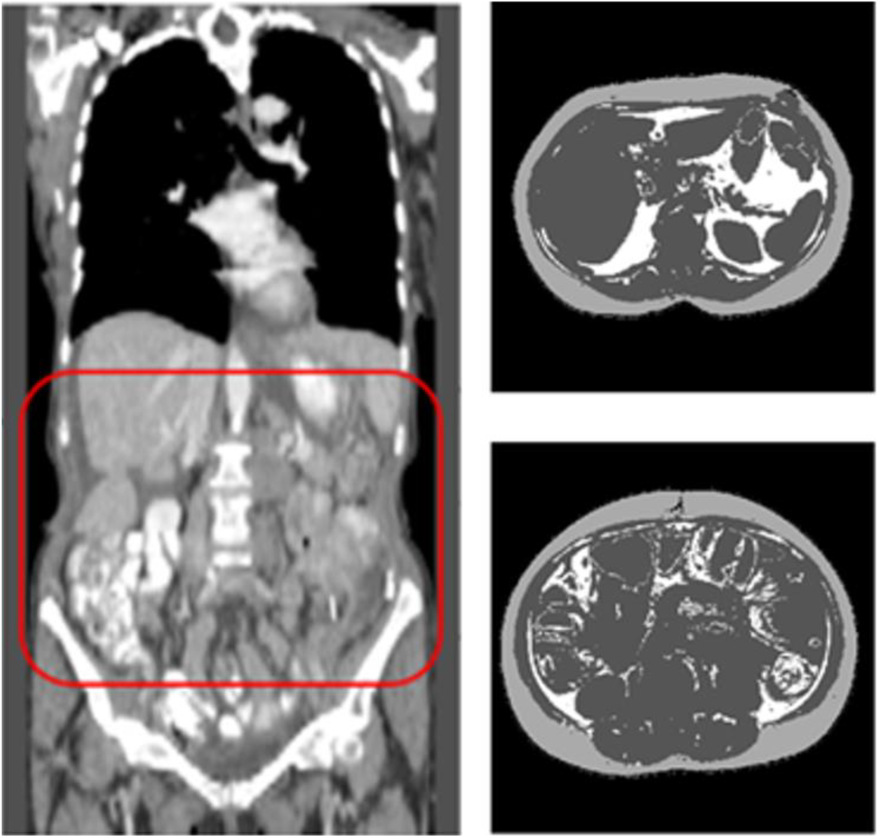
Fig. 1.

Illustration of measuring VFA and SFA from the CT images by applying a computer-aided detection scheme to automatically (a) detect abdominal section of CT images (inside a box with Red frame) and (b) segment each CT image slide into 3 categories (in which SFA is shown in light gray color, VFA in white color and other non-fat human organs in dark gray color).
