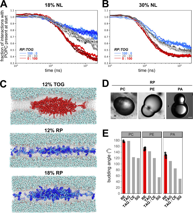
Figure 3.
Nucleation and budding properties of REs. (A and B) Coarse-grained MD simulations of lens formation by 150 (18%; A) and 250 (30%; B) neutral lipids (NL) per leaflet in a POPC membrane. Colors indicate the neutral lipid composition with marine blue for pure RE and red for pure TOG. The progress of lens formation is shown as fractional loss of interactions between the neutral lipids and POPC as a function of time (logarithmic scale). (C) Snapshots of systems with 12% TOG (top), 12% RP (middle), and 18% RP (bottom) obtained after backmapping and equilibrating the corresponding CG systems to charmm36 all-atom models. TOG carbon atoms are colored red, and oxygen atoms are colored pink. For RP, the retinyl moiety is colored bright blue, the palmitoyl group is colored marine blue, and the oxygen atoms are colored pink. (D) Brightfield microscopy images of DIBs of droplets containing neutral lipid RP and lipid surfactants PC, PE, or PA. Scale bars indicate 20 µm. (E) Comparison of quantified budding angles (mean ± SD) of RP-containing droplets with lipid surfactant PC (176.2 ± 2.8), PE (150.9 ± 4.0), or PA (129.5 ± 5.7) versus reported budding angles of TAG- and squalene (SQ)-containing droplets (Ben M’barek et al., 2017). Bar plot represents mean of at least eight individual measurements, which are shown as black dots.

