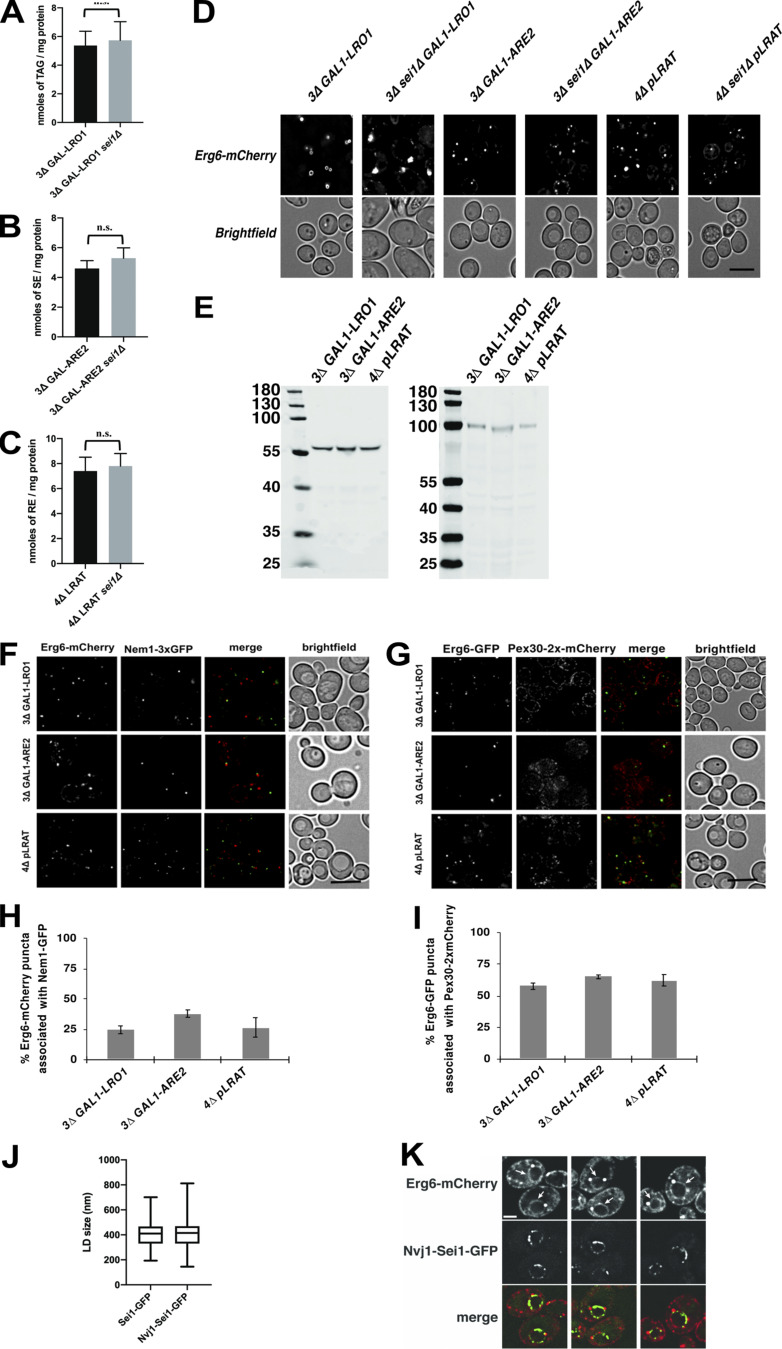
Figure S3.
Neutral lipids in cells with or without seipin. (A–C) Bar graphs showing TAG (A), SE (B), or RE (C) levels after induction with galactose (A and B) or ROH (C). Neutral lipids were measured with LC coupled to an ELSD. Cells were grown as described for Fig. 4 A; the amounts of neutral lipid indicate mean ± SD. Statistical significance was determined by Welch’s t test. ***, P < 0.001. (D) Fluorescence microscopy images of single neutral lipid strains with or without seipin expressing the LD marker Erg6-mCherry. Strains containing genes under the GAL1 promoter were grown in medium containing raffinose and galactose for 24 h and visualized live. Strains containing the plasmid expressing LRAT were grown in SC medium with ROH for 24 h. Single deconvolved images are shown from Z-stacks of 20 images. Scale bars indicate 5 µm. (E) The indicated strains containing plasmids that express Sei1-GFP or Nvj1-Sei1-GFP were grown as described in Fig. 5, A and B. Extracts were separated by SDS-PAGE and immunoblotted with anti-GFP antibodies. The expected molecular weights of the Sei1-GFP and Nvj1-Sei1-GFP fusions are 60 kD and 99 kD, respectively. Numbers are sizes of the molecular weight markers. (F–I) Association of nascent LDs with Nem1 (F and H) or Pex30 (F and H). Strains were grown and imaged as in Fig. 4 A. Scale bars indicate 5 µm. Bar graphs in H and I show mean ± SD from 100 cells from 3 independent experiments for the results shown in F and G, respectively. (J) A 3Δ sei1Δ GAL1-LRO1 strain expressing Erg6-mCherry and either Sei1-GFP or Nvj1-Sei1-GFP was grown in SC medium with raffinose; galactose was added to the medium; and, 4 h later, the cells were visualized by fluorescence microscopy. The size of 100 LDs (Erg6-mCherry signal) was determined from 3 independent experiments. There is no statistically significant difference between the samples using Student’s t test. Compare these results with GAL1-LRO1 strains in Fig. 4 B. (K) 3Δ GAL1-LRO1 cells expressing Erg6-mCherry and Nvj1-Sei1-GFP were grown at 30°C in SC medium with raffinose; galactose was added the medium; and, 4 h later, the cells were visualized. Erg6-mCherry is on both the ER and the surface of LDs. The nucleus is indicated with an arrow. Three examples are shown. Scale bar indicates 1 µm.

