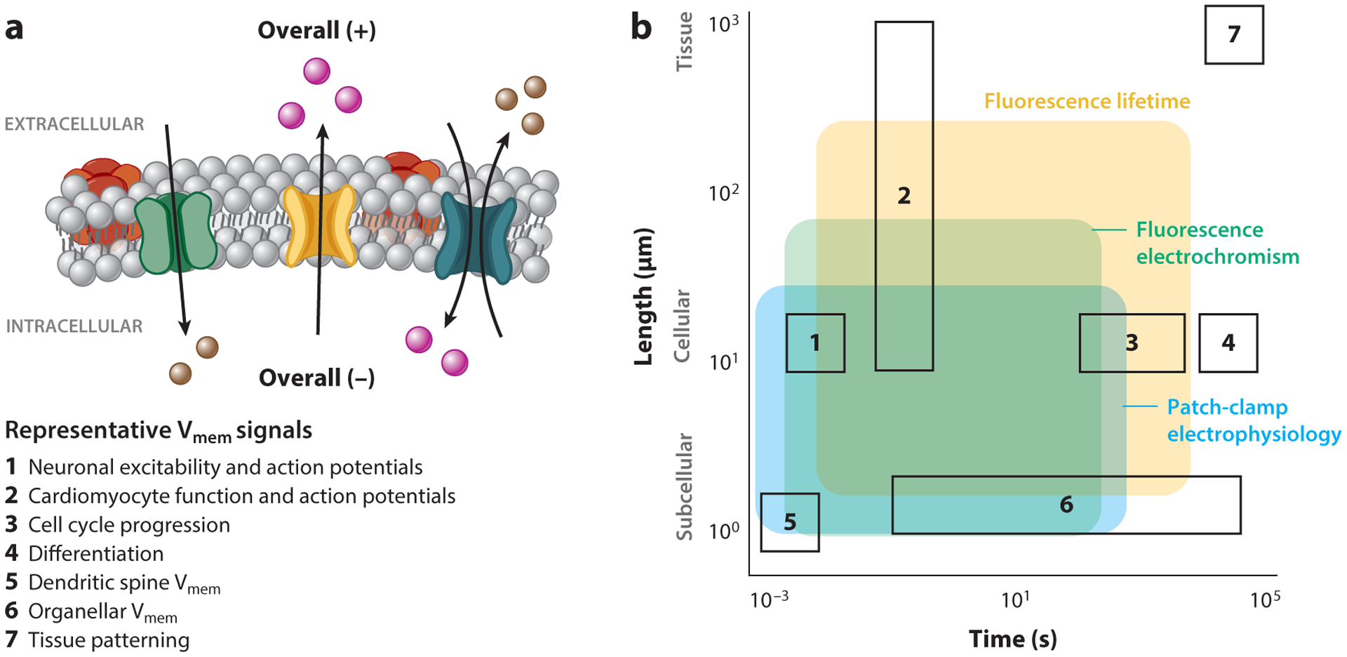
Figure 1.

Membrane potential (Vmem) signals and measurement techniques across time and space. (a) A schematic of cellular Vmem, with examples of seven biological processes where Vmem signaling plays a role. The resistance to ionic flow between compartments sets the length scale of Vmem signals; Vmem can scale across tissues or be compartmentalized within organelles or dendritic spines. The properties of ion channels and pumps that determine Vmem, as well as ion diffusion between compartments, dictate the time duration of a Vmem response. (b) Biological space accessible to the three most common strategies for absolute Vmem measurement (shaded regions), overlaid with example biological process of interest (outlined boxes, numbered as in panel a). While many techniques can report cellular Vmem on the scale of milliseconds or seconds, longer recordings or recordings across large areas are difficult to access. Shaded areas indicate regions where single trial recordings retain voltage resolution on the scale of biological Vmem changes (≤20 mV) and minimally alter the biological sample. Recordings with fluorescence electrochromic dyes require recalibration with an electrode on each cell, whereas fluorescence lifetime and patch-clamp electrophysiology can be applied as stand-alone measurements that are comparable between cells. Shaded areas are restricted to demonstrated application space for each technique and do not indicate the full extent of its possible use.
