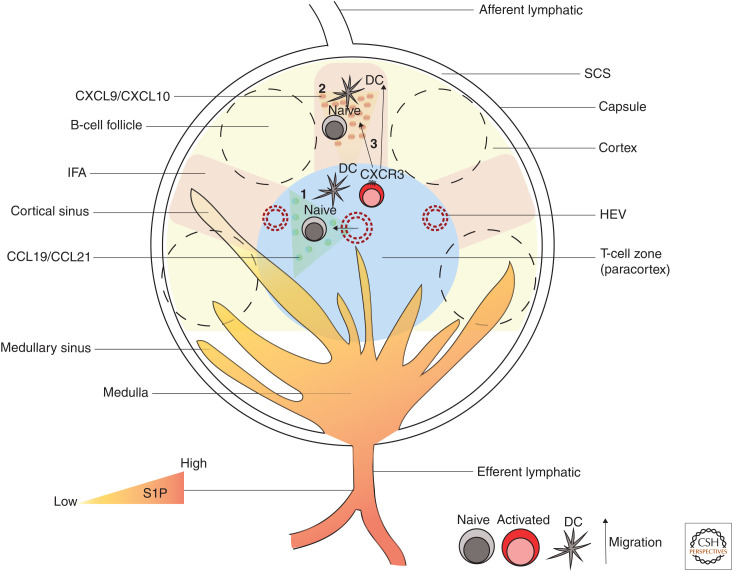
Figure 2.
Lymph node (LN) architecture and CD8+ T-cell priming. A schematic representation of the major regions of an LN and CD8+ T-cell priming sites. The main regions of LNs consist of the subcapsular sinus (SCS), B-cell follicles, interfollicular areas (IFAs), T-cell zone (paracortex), and the medulla. Lymph enters LN from afferent lymphatics and flows through the SCS. IFAs lie beneath the SCS and between B-cell follicles and are considered the inflammatory hotspots of LN as they contain innate immune cells that secrete various inflammatory molecules. Naive T cells enter LNs via high endothelial venules (HEVs) and locate to T-cell zones by responding to chemokines CCL19 and CCL21, where they get primed and activated by DCs (1). Alternatively, naive CD8+ T cells can also get activated in the IFA and SCS of LNs (2). Activated CD8+ T cells express CXCR3, which enables their migration toward the IFA, SCS guided by CXCL9 and CXCL10 gradients (3). Effector CD8+ T cells use sphingosine-1-phosphate (S1P) gradients in cortical and medullary sinuses to leave LNs via the efferent lymphatics.

