
Figure 2. Validation of WWS phenotype in vivo.
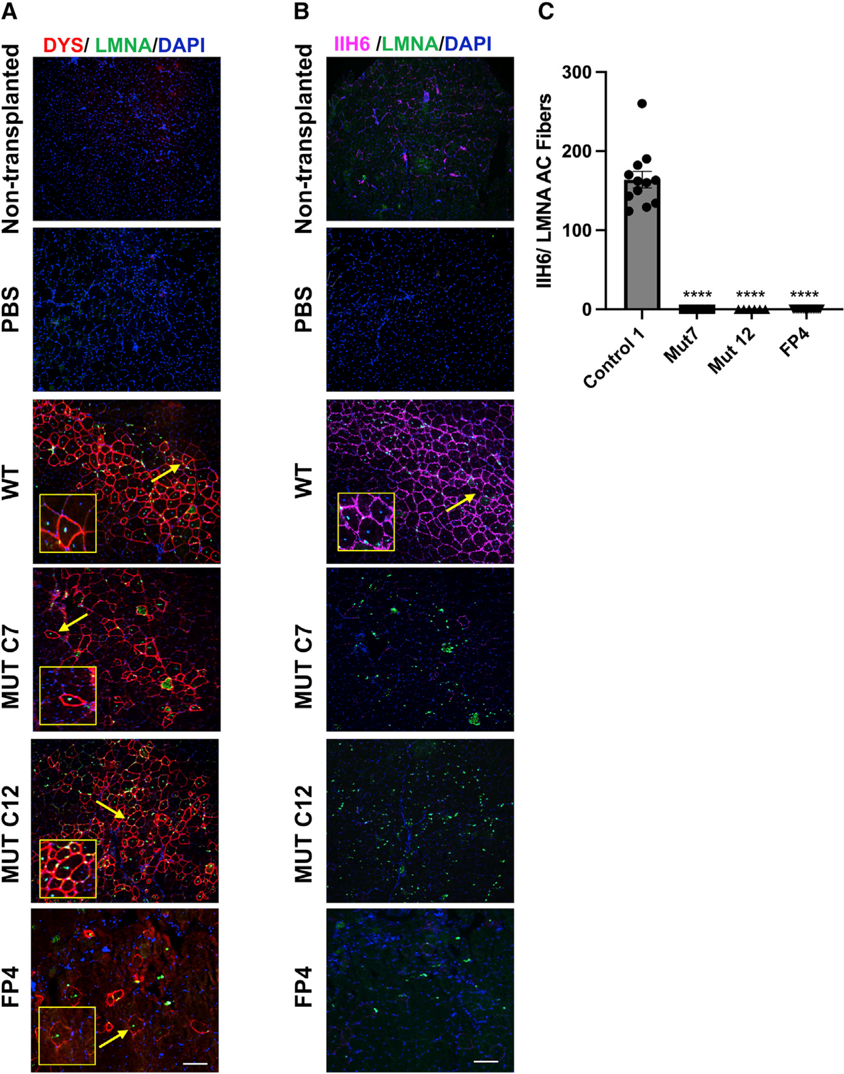
Representative images show the engraftment of myogenic progenitors derived from WWS FKRP mutant isogenic (WT and mutant clones C7 and C12) and FP4 patient-specific iPS cell lines upon their transplantation into TA muscles of FKRPP448L-NSG mice.
(A) Immunostaining for human DYSTROPHIN (DYS, in red) and human LAMIN A/C (LMNA, in green). DAPI in blue stains nuclei. PBS and non-transplanted muscle served as negative controls. Scale bar, 200 μm.
(B) Immunostaining for IIH6 (in purple) in combination with LMNA (in green). DAPI stains nuclei (in blue). PBS and non-transplanted muscle served as negative controls. Scale bar, 200 μm.
(C) Graph shows the quantification of the total number of donor-derived IIH6+/LMNA+ myofibers in TA muscles that had been transplanted with myogenic progenitors differentiated from control 1 (n = 12), MUT C7 (n = 4), MUT C12 (n = 4), and FP4 (n = 18) iPS cells. Data are shown as means ± SEMs. ****p < 0.0001 by ANOVA followed by the Tukey’s test.