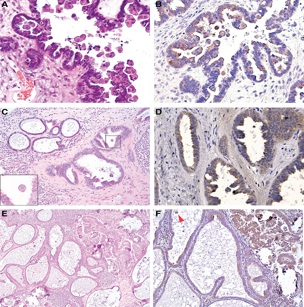
Figure 1.
Endosalpingiosis associated with BRAFV600E-mutated ovarian SBT (case #1). (A, B) Ovarian SBT, with cells exhibiting prominent eosinophilic cytoplasm, a histological feature associated with BRAFV600E mutation: (A) H&E (40×); (B) VE1 immunostaining (40×). (C, D) Associated nodal endosalpingiosis, composed of simple glands and adjacent glands with mild tufting and epithelial stratification: (C) H&E (4×), note scattered cells with dense eosinophilic cytoplasm (inset, 60×); (D) VE1 immunostaining (40×). (E, F) Lymph node involvement by SBT with adjacent endosalpingiosis: (E) H&E (10×); (F) VE1 immunostaining (20×). Arrowhead indicates positive staining in an exfoliated tumour cell with abundant cytoplasm.

