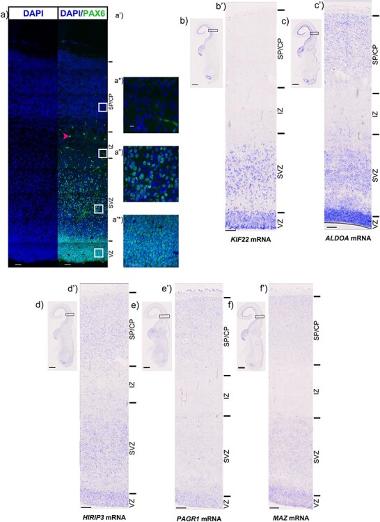
Figure 2 .

In situ hybridisation of candidate genes. (a) PAX6 protein (green) at 12pcw. a-a’*) show high magnification images of PAX6 protein expression in the (a’) SP/CP, a*) IZ, a”) SVZ and a’*) VZ. Low magnification scale bars = 50 μm, high magnification scale bars = 10 μm. b) Low magnification image of KIF22 mRNA in the 12pcw human fetal cortex, b’) High magnification showing KIF22 mRNA (blue) to be predominantly expressed in the germinative zones. c) Low magnification image of ALDOA mRNA in the 12pcw human fetal cortex, (b’) High magnification showing ALDOA mRNA (blue) to be predominantly expressed in the germinative zones but also some expression in the IZ and CP. (d) Low magnification image of HIRIP3 mRNA in the 12pcw human fetal cortex, (d’) High magnification showing HIRIP3 mRNA (blue) to be expressed throughout the telencephalic wall. e) Low magnification image of PAGR1 mRNA in the 12pcw human fetal cortex, e’) High magnification showing PAGR1 mRNA (blue) to be expressed throughout the telencephalic wall. (f) Low magnification image of MAZ mRNA in the 12pcw human fetal cortex, (f’) High magnification showing MAZ mRNA (blue) to be expressed throughout the telencephalic wall. For ISH, low magnification scale bars = 2 mm and high magnification scale bars = 100 μm.
