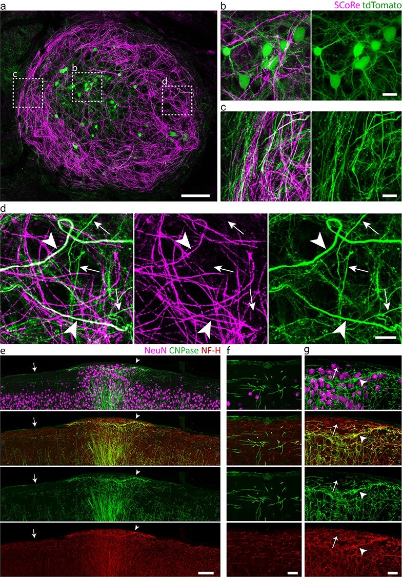
Figure 3 .

Myelinated and unmyelinated axons follow aberrantly looping and concentric paths. (a–d) Intravital imaging of a nest-like heterotopion at P30 in (a) using SCoRe and confocal fluorescence microscopy showing b, labeled neuronal cell bodies (tdTomato; green) dispersed throughout the aberrantly oriented myelinated fibers (SCoRe; magenta) and c, winding axonal projections that occasionally fasciculate into bundles at the edges of the heterotopion. (d) Myelinated (arrowheads) and unmyelinated axon segments (arrows) are observed within the heterotopion. (e–g) Low-magnification (e) and high-magnification (f, g) immunostainings of an embryonically induced heterotopion (g), confirming the presence of both myelinated (g, arrowhead) and unmyelinated (g, arrow) axon segments (f and g show high-magnification images of areas indicated by arrow and arrowhead in e, respectively). NF-H, neurofilament heavy chain. Images are representative of experiments performed in at least six animals. Scale bars, 100 μm (a), 15 μm (b–d), 100 μm (e), and 25 μm (f, g).
