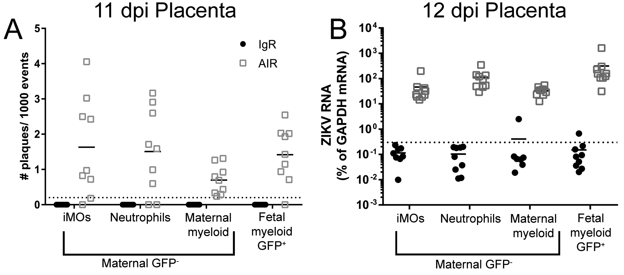
Fig. 2. Maternal and fetal myeloid cells in the placenta contain ZIKV RNA and infectious virus.

Placental immune cell enriched suspensions from 11 and 12dpi IgR and AIR GFP+ heterozygous litters were FACs sorted (Sup. Fig. 2) for myeloid cell populations. Infectious centers at 11dpi (A) or ZIKV RNA at 12dpi (B) for each sorted population from IgR (black symbols) or AIR (gray symbols) placentas was analyzed. Each dot represents sorted cells from a single placenta. Infectious centers are plotted as the number of plaques per 1000 events (cells) plated. Viral RNA was plotted as the percentage of gene expression relative to that of the Gapdh (glyceraldehyde 3-phosphate dehydrogenase) gene. The viral RNA level in each sample was calculated as the difference in the percentage in cycle threshold (Ct): Ct for Gapdh mRNA minus Ct for viral mRNA. The level of detection for the assay is indicated by the dotted line.
