Figure 5.
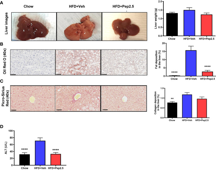
Peptide 19-2.5 abolishes steatohepatitis in HFD-fed mice. (A) Liver images of mice immediately after being culled and the liver weights (g). (B) Images of hepatic lipid deposition using Oil Red O staining and quantified as percentage of fat deposition (%). (C) Images of hepatic collagen deposition using Picro-Sirius Red stain to quantify percentage collagen deposition (%). Insert images are 40x magnification digital zoom and scale bars measure 50µm. (D) Serum ALT (U/L). Data were analyzed by one-way ANOVA, with Bonferroni’s post-hoc test. Data are expressed as mean ± SEM; n = 20 per group. **P < 0.01 and ****P < 0.0001 vs. HFD+Veh.

