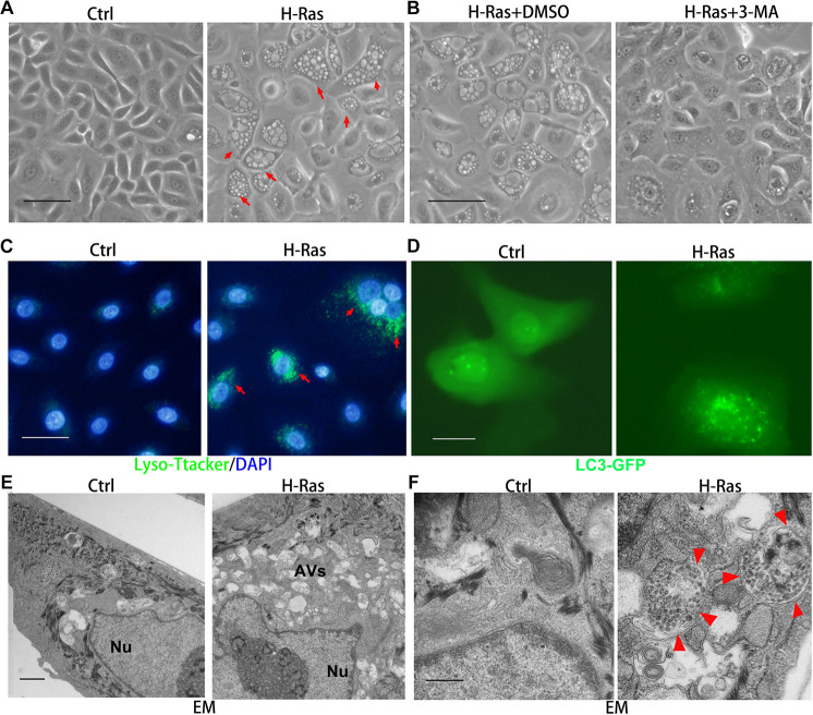
FIGURE 1.
H-Ras induces autophagic vacuoles in human keratinocytes. (A) Human keratinocytes were infected with a H-RasG12V expressing or a control retrovirus. Representative cell images 24 h after infection are shown, red arrows indicate cells with bubbles. (B) Representative images of human keratinocytes at 24 h after infection with a H-RasG12V expressing retrovirus plus treatment with DMSO as a control or with a PI3K inhibitor (3-MA). (C) Human keratinocytes were infected with a H-RasG12V expressing or a control retrovirus, at 24 h after infection, LysoTracker Green was added into the growth medium plus DAPI (blue for nuclei) for 5 min, then examined using a fluorescence microscope. (D) Human keratinocytes were transfected with a LC3-GFP expression vector, and after transfection, the cells were infected with a H-RasG12V expressing or a control retrovirus. At 24 h after infection, the cells were checked for GFP expression using a fluorescence microscope. (E,F) Human keratinocytes were infected with a H-RasG12V expressing or a control retrovirus. At 24 h after infection, the cells were processed for electron microscopic (EM) analysis. Low magnification images are shown in panel (E), and high magnification images are shown in panel (F). Nu: nuclei, AVs: autophagic vesicles, red arrows indicate the double membrane structure of autophagosomes. (A–D) Bars = 50 mm, (E) bar = 2 mm, and (F) bar = 100 nm.

