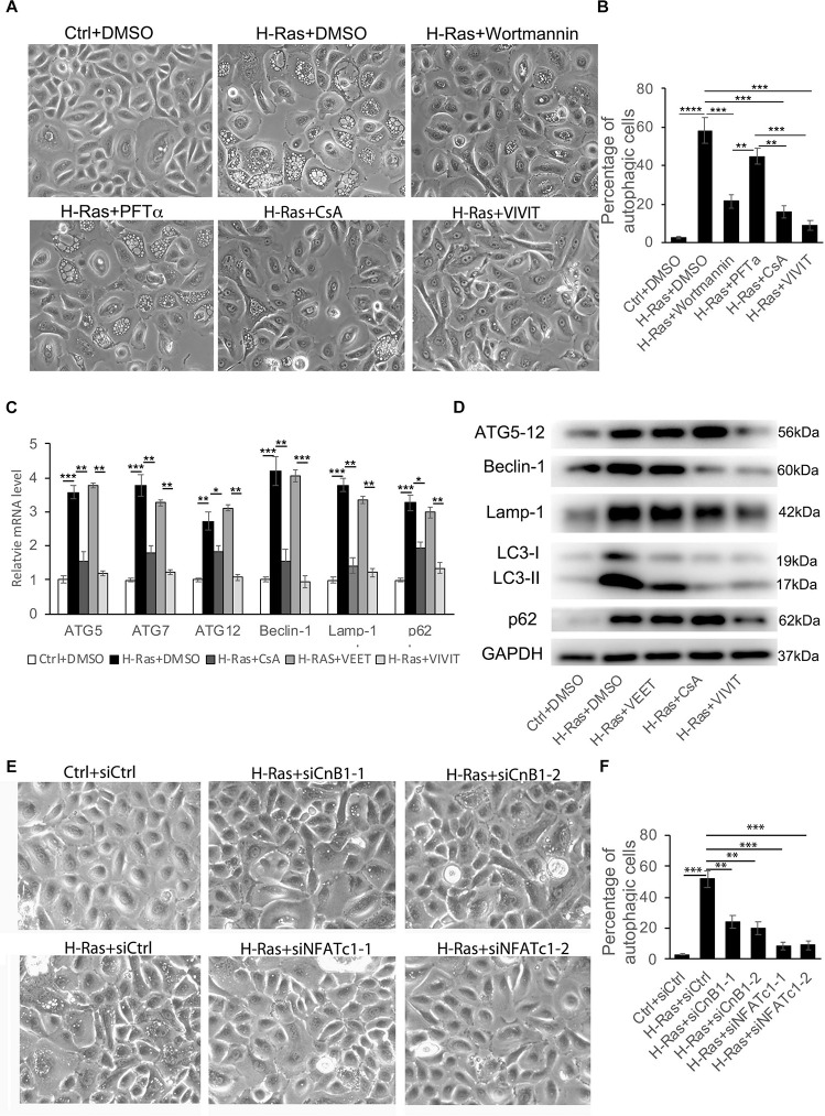
FIGURE 2.
H-Ras induced autophagy is suppressed by inhibition of the calcineurin/NFAT pathway. (A) Human keratinocytes infected with a H-RasG12V expressing or a control retrovirus were treated with different inhibitors as indicated, DMSO as a negative control. Representative cell images 24 h after treatment are shown. (B) Quantification analysis of the percentage of cells with bubbles from panel (A). (C) Human keratinocytes infected with a H-RasG12V expressing or a control retrovirus were treated with the calcineurin/NFAT inhibitors CsA or VIVIT, or with VEET as a control peptide for VIVIT for 24 h, then cells were collected for quantitative RT-PCR analysis of autophagy-associated genes as indicated. (D) Human keratinocytes were treated with the same conditions as shown in panel (C), then were lysed for immunoblotting analysis of autophagy-associated genes as indicated, GAPDH as a loading control. (E) Human keratinocytes were transfected with two independent siRNAs of CnB1 (siCnB1-1 and siCnB1-2) or NFATc1 (siNFATc1-1 and siNFATc1-2) or scramble siRNAs (siCtrl) as a control, and 2 days later, the cells were infected with a H-RasG12V expressing or a control retrovirus. Representative cell images 24 h after infection are shown. (F) Quantification analysis of the percentage of cells with bubbles from panel (E). (B,C,F) All experiments were carried out three times, and error bars represent means ± SD; p values are indicated with “*,” *p < 0.05, **p < 0.01, ***p < 0.005 when comparing two corresponding groups indicated with the black lines by Student’s t-test.

