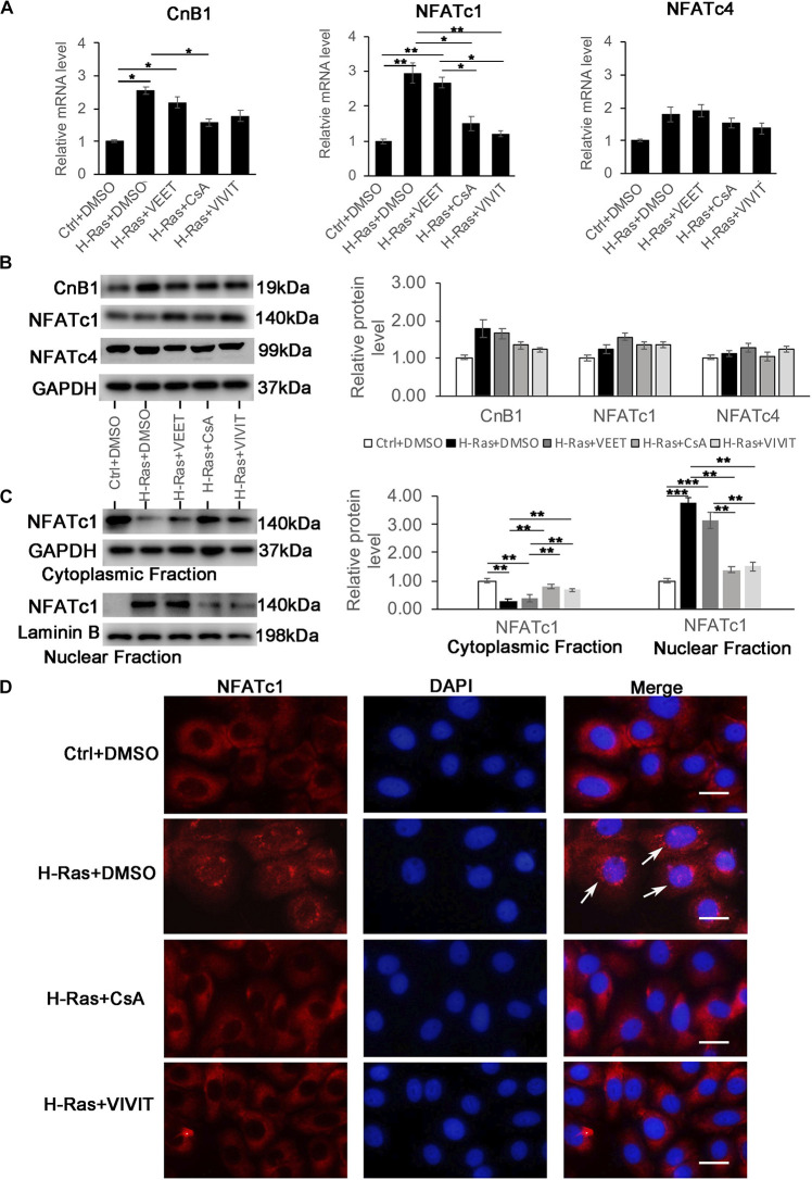
FIGURE 4.
Overexpression of H-Ras induces the nuclear translocation of NFATc1A. Human keratinocytes infected with a H-RasG12V expressing or a control retrovirus were treated with the calcineurin/NFAT inhibitors CsA, VIVIT, VEET as a control peptide for VIVIT or DSMO as negative control for 24 h, after which the cells were collected for quantitative RT-PCR analysis of calcineurin B1 (CnB1), NFATc1, and NFATc4 expression. (B) (Left panel) human keratinocytes were treated with same conditions as shown in (A), then cells were lysed for immunoblotting analysis of calcineurin B1 (CnB1), NFATc1, and NFATc4 expression, GAPDH as a loading control. (Right panel) Quantification of the relative levels of CnB1, NFATc1 and NFATc4 in the left panel. (C) (Left panel) human keratinocytes treated with same conditions as shown in panel (A) 24 h after infection, the cells were lysed for preparation of cytoplasmic and nuclear fractions. Both fractions were immunoblotted with the NFATc1 antibody, GAPDH as a loading control for the cytoplasmic fraction, and Laminin B as a loading control for the nuclear fraction. Right panel: Quantification of the relative levels of NFATc1 in the left panel. (A–C) All experiments were carried out three times, and error bars represent means + SD; p values are indicated with “*,” *p < 0.05, **p < 0.01, ***p < 0.005 when comparing two corresponding groups indicated with the black lines by Student’s t-test. (D) Human keratinocytes infected with a H-RasG12V expressing or a control retrovirus treated with the calcineurin/NFAT inhibitors CsA and VIVIT or DMSO as a control for 24 h, followed by immunofluorescence staining with the NAFTc1 antibody (red), and DAPI stain for nuclei. Merged images (Merge) are shown in the right column, and white arrows indicate the nuclear staining of NFATc1. Bars = 50 μm.

