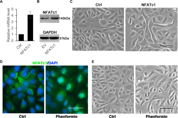
FIGURE 5.
Activation of NFATc1 doesn’t significantly induce vacuole formation in human keratinocytes. (A,B) Human keratinocytes were infected with a NFATc1 expressing or a control retrovirus, and 24 h after infection, the cells were collected for analysis of NFATc1 expression by RT-PCR (A) or by immunoblotting (B). GAPDH as a loading control in panel (B). The experiments were carried out three times, and error bars represent means ± SD; p values are indicated with “*,” **p < 0.01, when comparing with the control group by Student’s t-test in panel (A). (C) Representative images of human keratinocytes infected with a NFATc1 expressing or a control retrovirus for 24 h. (D,E) Human keratinocytes were treated with 1 mM phenformin for 24 h, then were fixed and analyzed NFATc1 (green) expression by immunofluorescence staining, DAPI stained for nuclei (blue). Representative images of human keratinocytes are shown in panel (E). (C–E) Bars = 50 mm.

