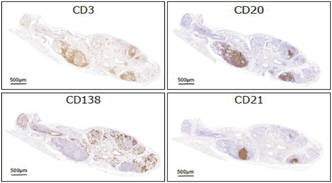
Fig. 1.
Immunohistochemical features of ectopic GCs in the SG of SS
Representative microphotographs depicting sequential immunohistochemistry staining for CD3 (T cells), CD20 (B cells), CD21 (follicular dendritic cell networks) and CD138 (plasma cells) identifying ectopic GC in labial salivary gland biopsy of SS patients. GC: germinal centres; SG: salivary glands.

