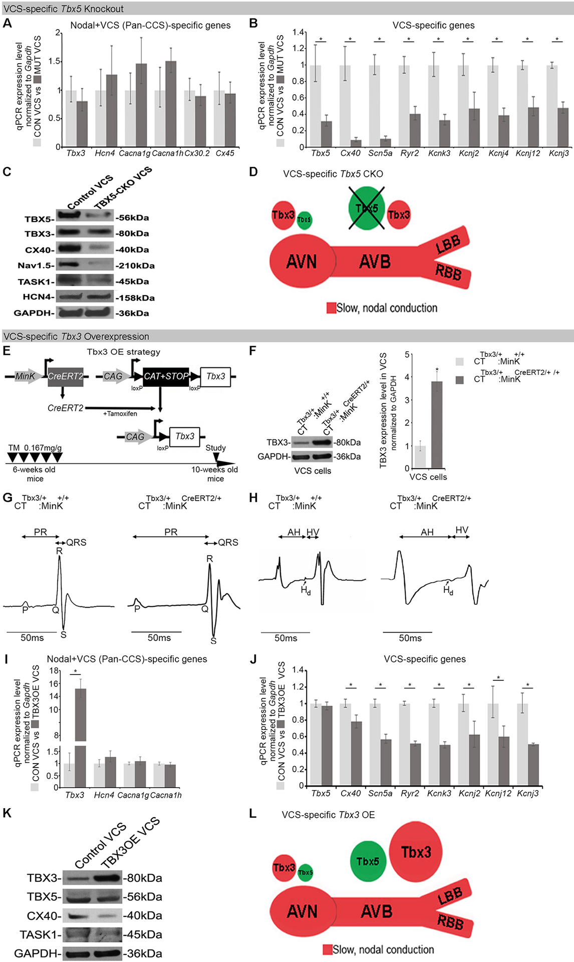
Figure 2. Proper Tbx3/Tbx5 balance is required to maintain expression of VCS-specific fast conduction genes.

(A-D) Characterization of Tbx5-dependent gene expression in the adult mouse VCS. Tbx5 was specifically removed from the mature VCS in Tbx5fl/fl;R26EYFP/+;MinKCreERT2/+ mice (7), TM treated at 6-weeks of age and analyzed by qRT-PCR with two sets of CCS-specific markers at 10 weeks of age. (A) One set of markers comprised genes normally expressed in both the node and VCS (Pan-CCS) and important for the slow conducting nodal phenotype. None of these genes demonstrated Tbx5-dependent VCS expression by qRT-PCR. (B) The second set comprised genes normally expressed specifically in the VCS (excluded from the nodes) and essential for VCS function. Expression of all these genes was profoundly Tbx5-dependent by qRT-PCR. (C) Immunoblotting analysis confirmed Tbx5-dependent expression in the VCS. (D) Graphical summary of transcriptional changes observed in VCS of VCS-specific Tbx5-deficient mice. Genetic deletion of Tbx5 from the mature VCS uncovers its slow conduction, nodal molecular phenotype. (E-L) VCS-specific TBX3 overexpression in adult CTTbx3/+;MinKCreERT2/+ mice. (E) Strategy to generate VCS-specific TBX3 overexpression in adult CTTbx3/+;MinKCreERT2/+ mice. CTTbx3 transgenic mice carrying a Cre-inducible TBX3 expression construct (CAG–CAT–Tbx3) (24) were crossed to VCS-specific tamoxifen inducible Cre transgenic mouse line (MinKCreERT2) (14) to generate CTTbx3/+;MinKCreERT2/+ mice. The control (CTTbx3/+:MinK+/+) and mutant (CTTbx3/+:MinKCreERT2/+) mice were tamoxifen administrated at 6 week of age. The effects of VCS-specific TBX3 overexpression were assessed 4 weeks after tamoxifen administration (at 10 weeks of age). (F) Western blotting analysis demonstrates 3.8 fold induced overexpression of TBX3 in the mutant VCS cardiomyocytes compared to their control littermates. (G,H) VCS-specific TBX3 overexpression causes significant VCS conduction slowing in adult CTTbx3/+;MinKCreERT2/+ mice. (G) Ambulatory telemetry ECG analysis showed increased PR and QRS intervals in CTTbx3/+:MinKCreERT2/+ mice compared to littermate CTTbx3/+;MinK+/+ control mice. Moreover, intracardiac electrophysiology detected increased A-H, H-V, and His duration (Hd) intervals (H), all indicative of VCS conduction slowing, only in CTTbx3/+:MinKCreERT2/+ mutants never in CTTbx3/+;MinK+/+ controls. (I,J) qRT-PCR analysis of molecular changes driven by Tbx3 overexpression in VCS of adult mice. VCS-specific Tbx3 overexpression did not affect expression of Pan-CCS-specific slow conduction genes (I), but caused a significant reduction in expression of VCS-specific fast conducting genes (J) in CTTbx3/+;MinKCreERT2/+ mutants compared to littermate CTTbx3/+;MinK+/+ controls. (K) Immunoblotting analysis confirmed a significant reduction in expression of VCS-specific fast conducting genes in CTTbx3/+;MinKCreERT2/+ mutants compared to littermate CTTbx3/+;MinK+/+ controls (L) Graphical summary of transcriptional changes observed in mice with induced VCS-specific Tbx3 overexpression. Data are presented as mean±SD. For (A, B, I, J), n=3 biological replicates/ genotype (VCS cardiomyocytes pooled from 30 mice per each biological replicate). For (C, F, K), n=2 biological replicates/ genotype (VCS cardiomyocytes pooled from 50 mice per each biological replicate). For (G), n=3 or 5 animals/ genotype. For (H), n=3 or 4 animals/ genotype. For (A, B, I, J), Welch’s t-test or Mann-Whitney U test, multiple testing correction using Benjamini & Hochberg procedure; *: FDR≤0.15. For (F-H), Welch’s t-test; *P<0.05. Abbreviations: OE, overexpression; A-H, Atrio-Hisian interval; H-V, Hisio-Ventricular interval; Hd, His-duration.
