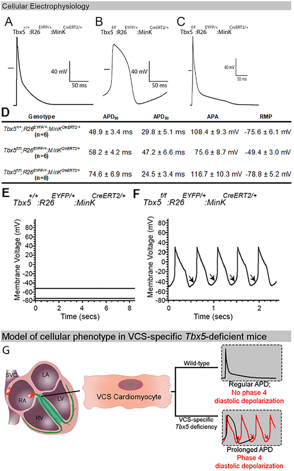
Figure 3. Electrophysiological characterization of mice with VCS-specific TBX5-deficiency.

(A-C) Action potentials (APs) from a control VCS (A), Tbx5-deficient VCS (B) and ventricular (C) cardiomyocyte by whole-cell patch–clamp. (A-B) AP of the Tbx5-deficient VCS cardiomyocytes demonstrate a slower upstroke (phase 0), longer plateau (phase 2), delayed repolarization (phase 3) and enhanced phase 4 depolarization compared to the control VCS cardiomyocyte. (C) AP of a ventricular cardiomyocyte from a VCS-specific, Tbx5-deficient mouse shows a rapid upstroke (phase 0), minimal plateau (phase 2), rapid repolarization (phase 3) and no phase 4 depolarization. The short horizontal bar in each panel represents the zero potential level. (D) Table showing detailed AP parameters recorded from mutant VCS, control VCS and ventricular cardiomyocytes. (E, F) VCS-specific removal of Tbx5 induces cellular automaticity in adult VCS cardiomyocytes. (E) No evidence of autonomous electrical activity, including phase 4 depolarization or autonomous beating, were observed in VCS cardiomyocytes isolated from control mice. (F) Phase 4 depolarization (arrows) and autonomous beating were observed in all VCS cardiomyocytes isolated from Tbx5-deficient mice. (G) Summary of cell autonomous defect observed in adult, Tbx5-deficient VCS cardiomyocytes. For (A-F), n=6 or 8 biological replicates/ genotype; Kruskal-Wallis H test; *P<0.05. Abbreviations: APD50, 90, AP duration at 50 and 90% of repolarization; APA, AP amplitude, RMP, resting membrane potential.
