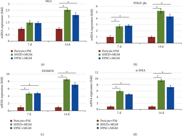
Figure 6.

The expression of pericyte-specific cell surface markers after culture in Pericyte Medium (PM) and Melanocyte Growth Medium (MGM)for 14 d. (a) Stem cells from human exfoliated deciduous teeth (SHEDs) (NG2+, 23.3%; DESMIN+, 83.7%; PDGFR-β, 93.3%; α-SMA, 93.3%). (b) Pericytes (NG2+,74.3%; DESMIN+,67.8%; PDGF-β, 85.6%; α-SMA, 92.3%). (c) Postnatal human dental pulp stem cells (DPSCs) (NG2+,15.1%; DESMIN+, 58.4%; PDGFR-β, 80.6%; α-SMA, 91.4%).
