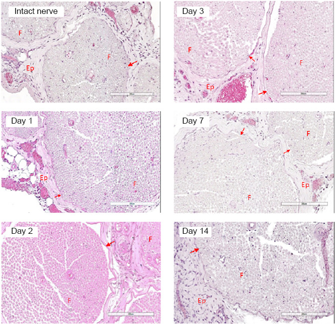
Figure 2.

Cross sections of rat sciatic nerve 1, 2, 3, 7, and 14 days after intraneural administration of the plasmid pBud-coVEGF165-coFGF2.
Hematoxylin and eosin staining. No inflammatory or degenerative features observed. Ep: Epineurium; F: fascicle. Red arrows correspond to perineurium. Scale bars: 200 μm. Intact nerve refers to nerve injected with phosphate buffer saline.
