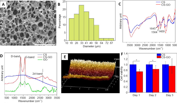
Figure 1.

Analyses of the structure, composition, and properties of the CS-GO scaffold.
(A) Scanning electron microscope image of the CS-GO scaffold. The pores of the CS-GO scaffolds were irregular and randomly distributed. Scale bar: 200 μm. (B) Distribution of the micropore diameters in the GS-GO scaffold. (C) Fourier-transform infrared spectroscopy spectra and (D) Raman spectra of CS and CS-GO scaffolds. Amide I band of C=O stretching at 1645 cm‒1, amide II band of C-N stretching at 1564 cm‒1, and amide III band of N-H deformation at 1409 cm‒1 peaks were observed in the CS and CS-GO scaffolds. There was a distinct D-band at 1350 cm‒1, G-band at 1605 cm‒1, and 2D-band at 2657 cm‒1 in the pattern of CS-GO. (E) Atomic force microscope image of the surface of the CS pores. (F) 3-(4,5-Dimethylthiazol-2-yl)-2,5-diphenyltetrazolium bromide results of PC12 cells cultured on CS and CS-GO scaffolds. The cell viability was expressed as the optical density ratio to the CS group at day 1. Data are expressed as mean ± SEM. *P < 0.05 (two-tailed Student’s t-test). CS: Chitosan; CS-GO: graphene oxide-composited chitosan; GO: graphene oxide
