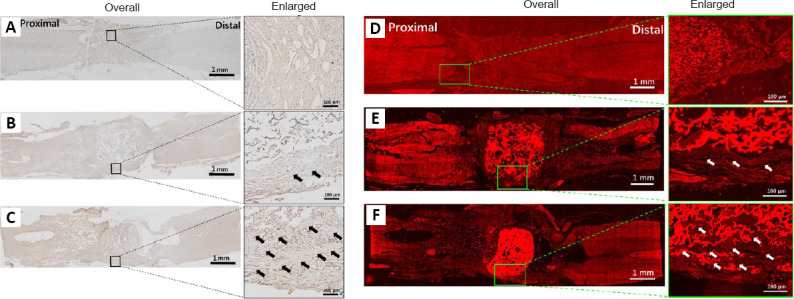
Figure 4.

Evaluation of different stained repair tissue of the injured site 10 weeks after SCI.
(A–C) Evaluation of tissue remodeling by immunohistochemistry and (D–F) immunofluorescence (ChAT). (A, D) Control, (B, E) CS, and (C, F) CS-GO groups. There are a lot of interval spaces in the Control and CS scaffold groups; however, the repair tissue grew into the gaps of the CS-GO group. ChAT (a neuronal marker; red, stained by Alexa Fluor 568). The arrows point to regenerated neural tissue. Scale bars: 1 mm (left) and 100 μm (right). ChAT: Choline acetyltransferase; CS: chitosan; CS-GO: graphene oxide-composited chitosan; GO: graphene oxide.
