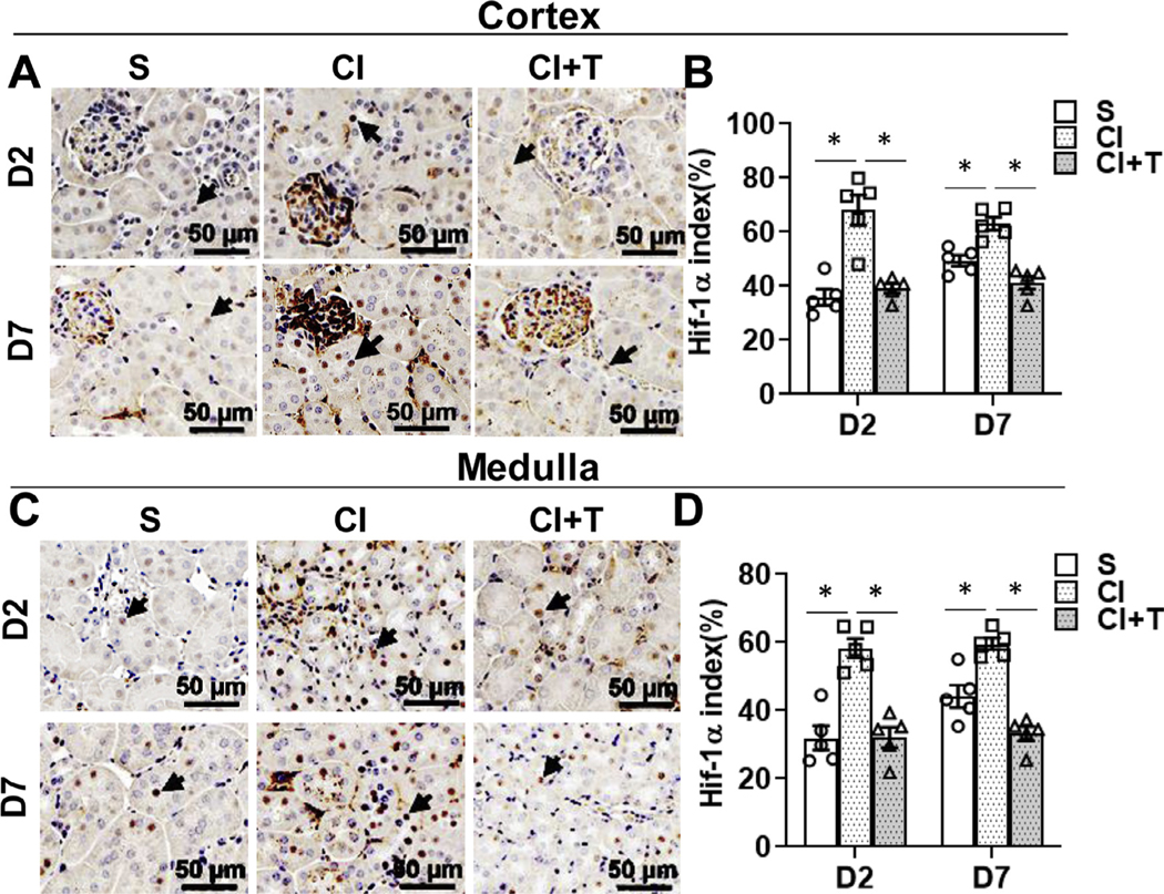
Fig 6.

Terazosin treatment abrogates contrast-induced hypoxic response in kidneys. Hypoxia inducible factor-1α (HIF-1α) was assessed by immunohistochemistry (A and C) in mouse kidneys at day 2 (D2) and day 7 (D7) after contrast (CI) or saline (S) administration. Representative Immunohistochemistry images of HIF-1α protein staining in the cortex (A) and in medulla (C) at D2 and D7 of contrast administration. The arrow heads point to brown HIF-1α positive nucleus in the tubular cells. The intensity of brown staining was quantified in cortex (B) and medulla (D) using Zen Pro image analysis software and presented as bar graph. S, mice with saline administration IP; CI, mice with contrast administration; CI + T, mice with contrast administration plus terazosin IP injection. ANOVA with repeated measurements was performed with post hoc Student t-test. Each bar represents mean ± SEM of 5 animals per group. * indicates P < 0.05.
