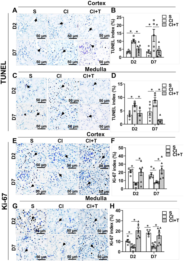
Fig 7.

Terazosin treatment abrogates contrast-induced cell death and increases cell proliferation in kidneys. Cell death and cell proliferation were assessed by cells positive for TUNEL staining (apoptosis) and immune staining for Ki-67, respectively, in kidneys of mice at day 2 (D2) and day 7 (D7) after contrast administration. Representative images of TUNEL in cortex (A) and in medulla (C), and Ki-67 in cortex (E) and in medulla (G) staining at D2 and D7 of contrast administration. The arrow head points to brown positive nuclei for TUNEL (A and C) and Ki-67 (E and G) staining. The percentage of TUNEL or Ki-67 positive nuclei were counted using Zen Pro image analysis software. S, mice with saline administration IP; CI, mice with contrast administration; CI + T, mice with contrast administration plus terazosin IP injection. ANOVA with repeated measurements was performed with post hoc Student t-test. Each bar represents mean ± SEM of 5–7 animals per group. * indicates P < 0.05.
