Figure 2.
Functions of Membrane-Associated Mucins at the Human Ocular Surface.
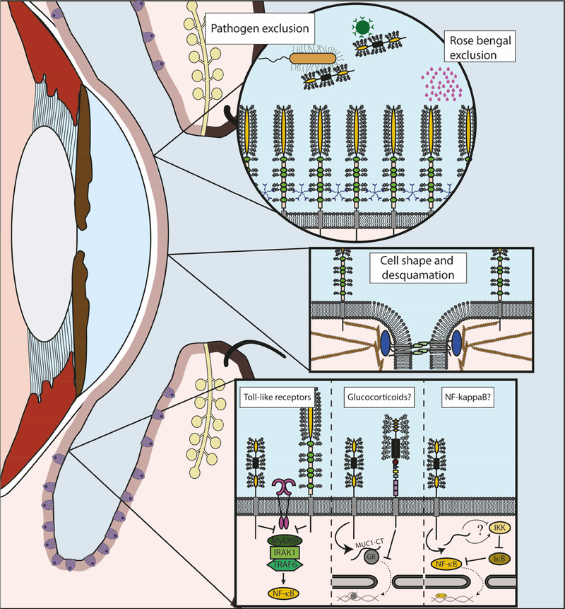
The graphic depicts some of the focus areas of this article.
Left. Sagittal section of the eye and eyelids is represented, with epithelia highlighted in light pink. Goblet cells can be observed in the conjunctival epithelium (purple). Meibomian glands (yellow) can be found at the lids, with their ducts opening at the lid margin.
Top right. Detail of the barrier functions of MAMs. Multiple MUC16 are showed protruding from the plasma membrane as the main elements of the glycocalyx barrier. LGALS3 (galectin-3) pentamers can be found interacting with mucin glycans. This barrier is responsible of the exclusion of different substances, such as the clinical dye rose bengal. Bacteria and viruses can be excluded also by shed mucins, as it is the case of MUC1, represented here.
Mid right. Detail of the interactions of MAMs with elements of the cytoskeleton. MUC16 participates in the formation of membrane microplicae and tight junctions (represented here with the transmembrane protein occluding and ZO1 in blue). Some of these functions have been related to their interaction with actin filaments.
Bottom right. Detail of the immunomodulatory functions of MAMs. A question mark is used to indicate functions that have not been demonstrated in the eye to date. Inhibitory effect on Toll-like receptors (fuchsia) has been described for MUC1 (left) and MUC16 (right) at the ocular surface, reducing the activation of NF-kappaB. Previous studies showed that MUC1 acted by inhibiting recruitment of MyD88. Studies in airways have demonstrated that MUC1-CT interacts with the glucocorticoid receptor (NR3C1), facilitating its migration to the nucleus, while MUC4 (right) can inhibit its translocation. Finally, studies in different systems have described interactions between MUC1-CT and different regulatory elements of the NF-kappaB pathway, although the final result of these interactions is not clear.

