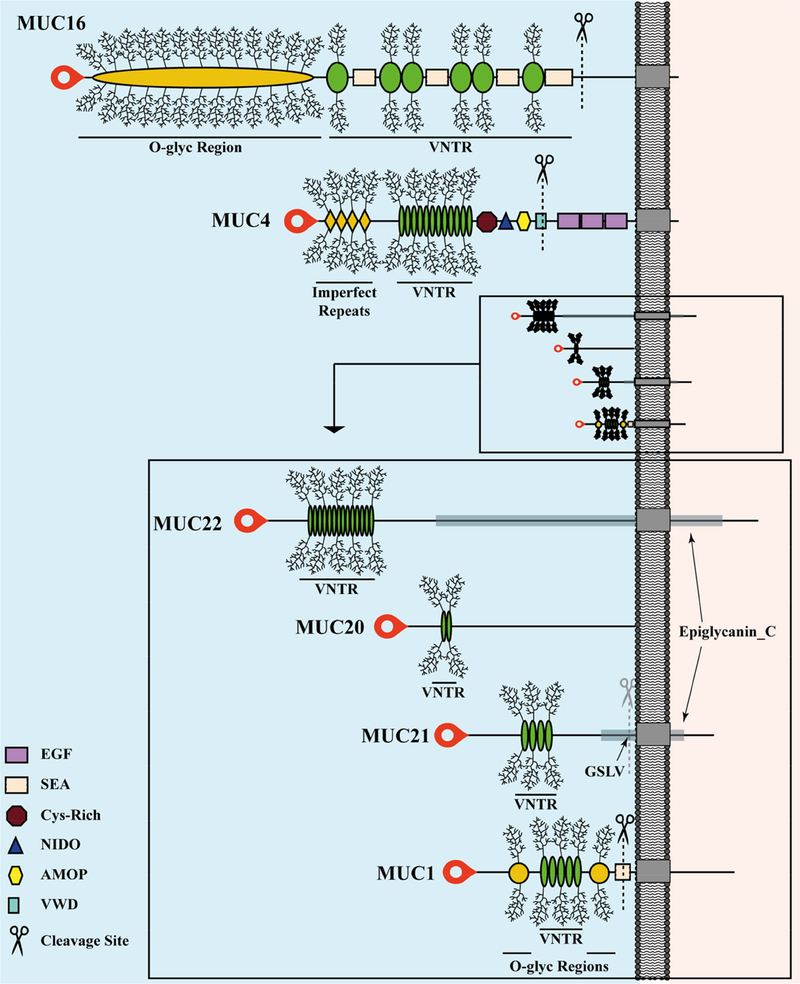
Figure 3.
Modular Architecture of Membrane-Associated Mucins of the Human Ocular Surface.
The extended conformation of each extracellular domain (ED) is to the left of the plasma membrane (gray bar) and each cytoplasmic tail (CT) to the right, both drawn to scale. The transmembrane domains are indicated as gray boxes embedded in the plasma membrane. MUC20 has been experimentally determined to associate with the plasma membrane, but has no transmembrane domain, and thus no CT has been identified (discussed in the text). Because of the extreme size differences between the large and small MAMs, an expanded view of the ED is shown for the small MAMs. Signal peptides are located at the amino-terminus of each protein (red blobs). The cleavage sites in the EDs are indicated by scissors.

