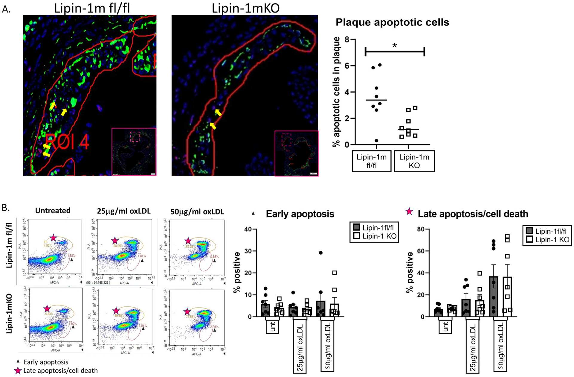
Fig. 3.

Lipin-1mKO mice have fewer apoptotic cells in plaques.
(A) Aortic root tissue sections from wild-type and lipin-1mKO mice on high fat diet for 12 weeks were stained for apoptotic cells, macrophages, and DAPI. Apoptotic cells within the plaques, shown with yellow arrows, were quantified as cells positive for DAPI (blue) and TUNEL (red) (B) Wild type and lipin- KO bone marrow-derived macrophages were treated with oxLDL for 24hrs. Cells were then stained with Annexin V and PI and analyzed via flow cytometry. Early apoptosis = Annexin V + and PI−. Late apoptosis = Annexin V+ and PI+. *p < 0.5 Student t-test analysis was used for statistical analysis between wild type and lipin-1mKO.
