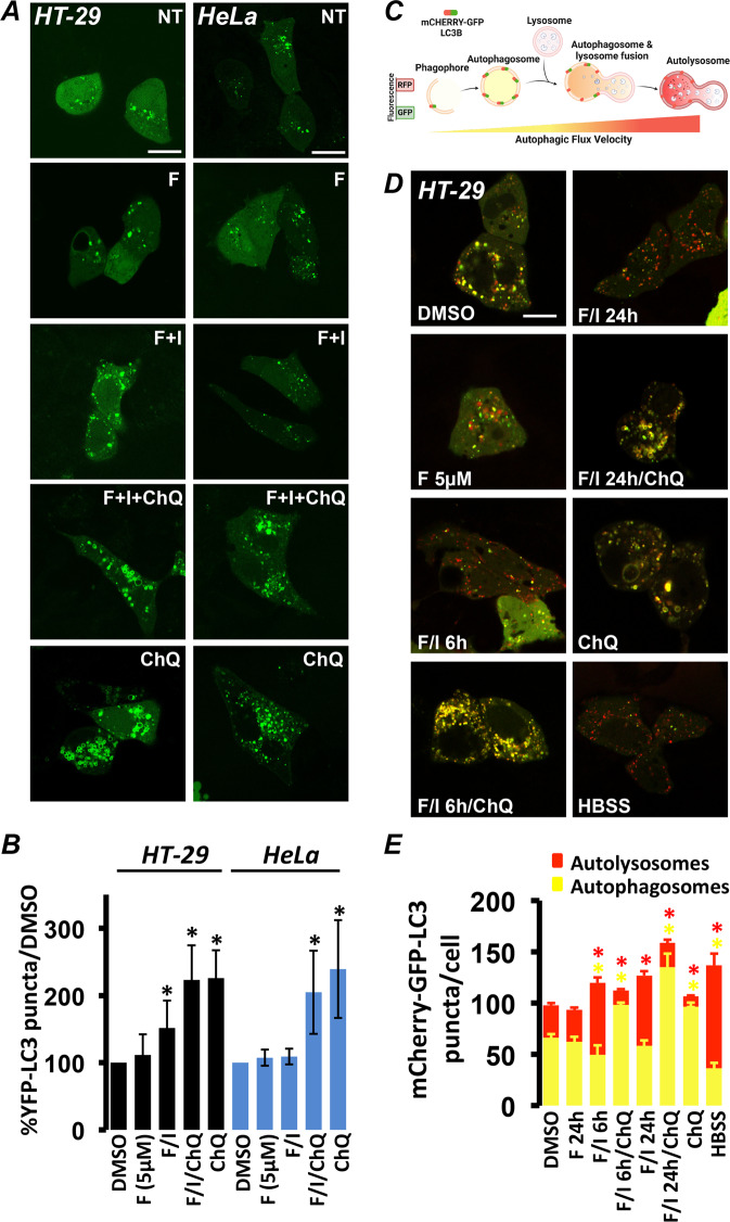
Fig. 1. Cyclic AMP elevating agents increase the autophagic flux in HT-29 but not HeLa cells.
A Confocal photomicrographs of HT-29 and HeLa cells expressing YFP-LC3. Treatment with 5 µM forskolin (F) did not alter the number of YFP-LC3-positive structures, while 20 µM F combined to 500 µM IBMX (F/I) increased YFP-LC3 puncta in HT-29 but not in HeLa cells. Chloroquine (ChQ) (100 µM) was used as control. B Summary of the effects on YFP-LC3 puncta of each treatment normalized to vehicle control (DMSO). Average of HT-29 cells: DMSO: 282, F 5 µM: 77, F/I: 276, F/I/ChQ: 31, ChQ: 38 in at least six independent experiments. Average of HeLa cells: DMSO: 98, F 5 µM: 58, F/I: 80, F/I/ChQ: 69, ChQ: 67 in at least four independent experiments. (*p < 0,04; **p < 0,01). C Schematic representation of how the sensor mCherry-GFP-LC3 works (created with BioRender.com). D Confocal photomicrographs of HT-29 cells expressing mCherry-GFP-LC3. Treatment with 5 µM forskolin (F) did not alter either the number or the balance between autophagosomes (mCherry+/GFP+) and autolysosomes (mCherry+/GFP–). Treatment with 20 µM F combined to 500 µM IBMX (F/I) for 6 or 24 h increased the number of autolysosomes and decreased the number of autophagosomes similarly to starvation (HBSS). Treatment with ChQ alone or in combination to F/I drastically decreased the number of autolysosomes and increased the number of autophagosomes. E Summary of the effects on mCherry-GFP-LC3 puncta number and type of each treatment. Average of HT-29 cells: DMSO: 83, F 5 µM: 38, F/I/6 h: 31, F/I/24 h: 37, F/I/6 h/ChQ: 12, F/I/24 h/ChQ: 15, ChQ: 40, HBSS: 22 in at least three independent experiments (*p < 0,01) (Scale bar 20 µm).

