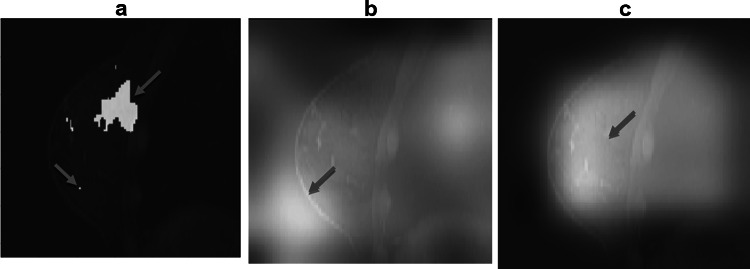
Fig. 3.
a Shows the tumor mask, extra pixels, e.g., one in the left lower quadrant, are present because the way segmentation is performed from the difference image between contrast-enhanced and no-contrast MRI, b visualizes that the class activation map of no-mask model 2 is off the breast tissue, and c visualizes that the class activation map of mask-guided model 3 finds the regions of interest inside the breast but outside the mask

