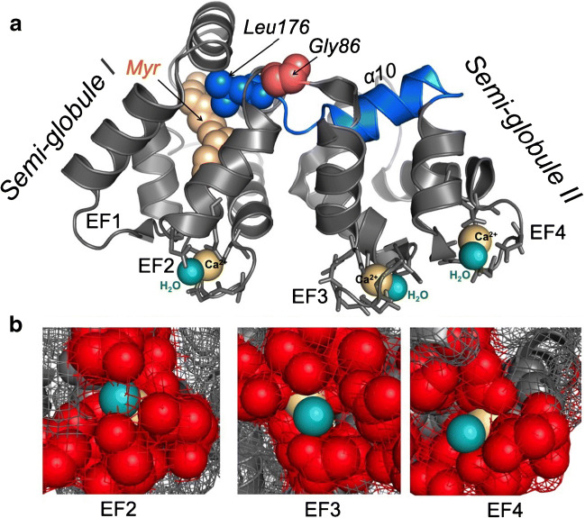
Fig. 4.
a Three-dimensional ribbon diagram of GCAP1 indicates the positions of Ca2+ (yellow) and H2O (light cyan) coordinated in three EF-hand loops; the calcium-myristoyl tug structure is highlighted in blue and the hinge Gly-86 between the two semi-globules is shown in pink. b Space-filled/mesh close-up diagram of the Ca2+:H2O position in EF-hand structures; red space-filled side chains in the loop, yellow Ca2+, cyan H2O (the models utilize the molecular coordinates of the GCAP1 crystal structure reported by Stephen et al. [111]

