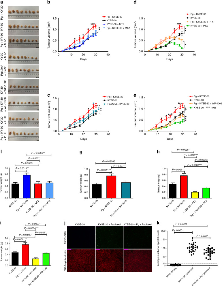
Fig. 5. Porphyromonas gingivalis infection aggravates progression of ESCC through STAT3 signalling in a xenograft tumour-bearing model.
KYSE-30 cells were pre-treated with wild-type P. gingivalis (MOI 10) or P. gingivalis ΔfimA mutant for 24 h and implanted in athymic nude mice (n = 8). Tumour cells were inoculated with the same amount of P. gingivalis at days 10 and 15. Metronidazole (30 mg/kg), WP1066 (20 mg/Kg), paclitaxel (10 mg/kg), and solvent control were administered as described in “Methods”. a Representative tumour specimens dissected from the athymic nude mice xenografted with KYSE-30 cells with different treatments at the end of the study. The average tumour volume (b–e) and tumour weight (f–i) were calculated at the time indicated. As compared with the control group, infection of P. gingivalis significantly promoted tumour growth (a), while loss of FimA (c, g) or metronidazole treatment (b, f) abrogated the pro-tumorigenic ability of P. gingivalis in KYSE-30 inoculated athymic nude mice. Upon treatment with paclitaxel (d, h) or WP1066 (e, i), the volume and weight of xenograft tumours were significantly decreased in mice inoculated with P. gingivalis-infected KYSE-30. Infection with P. gingivalis also significantly reduced the paclitaxel-induced apoptosis in xenograft tumour tissue from mice (j, k). Representative images (j) and an average number (k) of apoptotic cells. The data in (j and k) represent the mean ± standard deviation of apoptotic cells from three non-consecutive tissue sections of each mouse. All results are the average of at least three independent experiments. Error bars represent standard deviation. *, **, and *** represent P < 0.05, P < 0.01, and P < 0.001, respectively.

